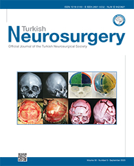
MATERIAL and METHODS: A total of 31 infants with premature closure of coronal suture were randomly divided into two groups; experimental group (n=16) and control group (n=15). In the experimental group, the skull model was reconstructed by an imaging examination and three-dimensional (3D) printing technique before the operation, and the fronto-orbital band was anteriorly displaced during the operation to guide the surgical treatment of craniosynostosis. In the control group, the skull model was reconstructed by an imaging examination and 3D printing technique before the operation, and the fronto-orbital band was not anteriorly displaced during the operation by the same operator. The surgical effects of the two groups were compared.
RESULTS: During the 12-month follow up after the operation, the cephalic index of short head deformity in the experimental group was 80.7 ± 1.1, while that in the control group was 89.3 ± 4.5. There was a significant difference between the two groups.
CONCLUSION: Fronto-orbital band anterior displacement may guide the operation of craniosynostosis and significantly improve the effectiveness of surgical treatment of children with premature closure of coronal suture, which is worth popularizing in the clinical management of cases.
Keywords : Infant, Craniosynostosis, Surgery, Cranial suture, 3D printing technique


